A biology laboratory manual is a comprehensive guide for conducting experiments, offering detailed procedures, safety protocols, and essential techniques to explore biological concepts through hands-on learning experiences.
1.1 Importance of Laboratory Work in Biology
Lab work is integral to understanding biological concepts, enabling students to explore phenomena firsthand. It fosters critical thinking, observation, and experimental skills, enhancing scientific literacy. Hands-on activities bridge theory and practice, making complex ideas tangible. Through experiments, students develop analytical and problem-solving abilities, essential for scientific inquiry. Lab experiences also cultivate teamwork, data interpretation, and technical proficiency, preparing learners for real-world applications in biology. By engaging in structured investigations, students build a deeper appreciation for biological processes and their relevance to everyday life, fostering a stronger foundation for advanced studies and careers in science.
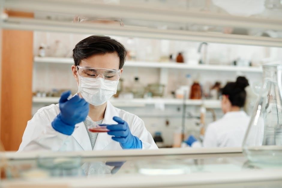
1.2 Overview of Laboratory Safety and Precautions
Laboratory safety is crucial to prevent accidents and ensure a secure environment for students and researchers. Key precautions include wearing personal protective equipment (PPE) such as gloves, goggles, and lab coats. Students must understand proper handling of biological specimens, chemicals, and equipment to minimize risks. Safe disposal of hazardous materials and adherence to emergency protocols, like fire evacuation plans, are essential. Clear communication and following instructor guidelines help maintain a safe workspace. Regular training and awareness of potential hazards ensure a protected learning environment, fostering responsible and confident lab practices for all participants.
Essential Laboratory Techniques
Mastering microscope usage, cell staining, and DNA extraction are fundamental skills. These techniques, along with PCR and protein analysis, form the backbone of molecular biology practices.
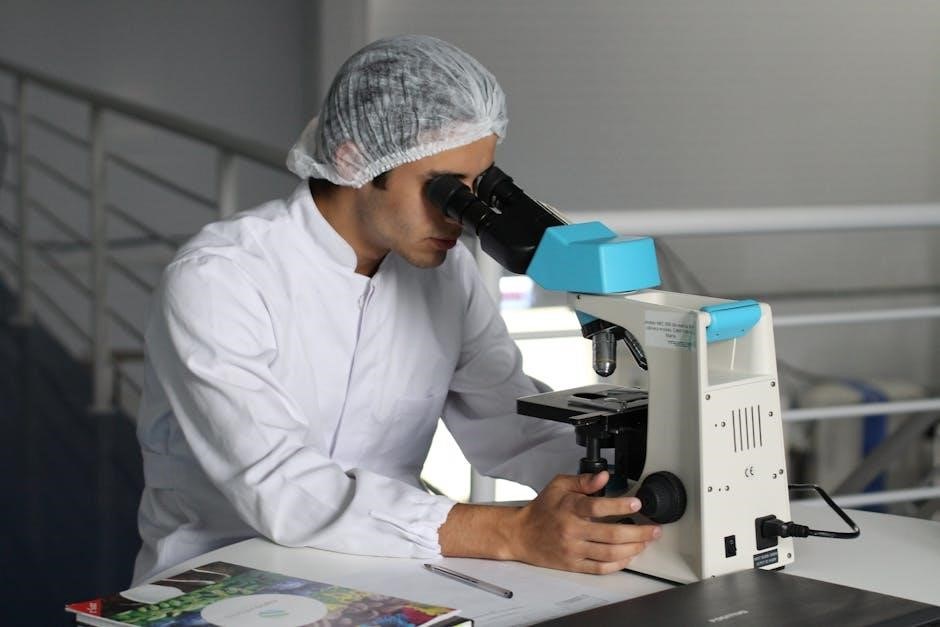
2.1 Microscope Usage and Maintenance
Microscope usage is fundamental in biology for observing microscopic structures. Proper preparation involves cleaning lenses with soft cloth and distilled water. For high-resolution imaging, immersion oil is applied between the objective and slide. Maintenance includes storing the microscope in a dry place, ensuring all parts are secure, and regularly checking for dust or debris. Familiarity with focusing mechanisms and stage movement enhances efficiency. Troubleshooting common issues like blurry images or mechanical malfunctions is essential for optimal performance. Regular maintenance extends the microscope’s lifespan and ensures accurate observations in various biological experiments.
2.2 Cell Structure and Staining Methods
Understanding cell structure is enhanced through staining techniques, which improve visibility of cellular components under a microscope. Common methods include Gram staining for bacteria and the use of dyes like methylene blue and safranin to differentiate cell parts. Fixatives such as formalin preserve cell structures before staining; Safety precautions are essential due to potential chemical hazards. Staining connects to broader biological concepts, aiding in the exploration of cell function and their roles in organisms. This practical approach reinforces theoretical knowledge, making cell biology accessible and engaging for students. Proper handling and disposal of chemicals ensure a safe learning environment.
Experiments in Cellular Biology
This section explores fundamental cellular processes through experiments such as diffusion and osmosis, and observing mitosis in plant cells. These activities provide practical insights into cellular functions and structures, reinforcing theoretical concepts and promoting a deeper understanding of biological mechanisms.
3.1 Diffusion and Osmosis Studies
Diffusion and osmosis are essential processes in cellular biology, enabling the transport of molecules across cell membranes. Lab experiments, such as those involving dialysis tubing or potato cells, demonstrate these phenomena. Students observe how substances move from areas of high to low concentration, understanding passive transport mechanisms. These studies highlight the role of concentration gradients and membrane selectivity. By analyzing results, learners gain insights into how cells maintain homeostasis and respond to environmental changes. Such experiments are fundamental for grasping cellular physiology and preparing for advanced topics in molecular biology.
3.2 Observing Mitosis in Plant Cells
Studying mitosis in plant cells provides a clear view of the cell division process. Students prepare specimens, such as onion root tips, by fixing and staining them to observe chromosomal changes. Using microscopes, they identify stages like interphase, prophase, metaphase, anaphase, and telophase. This exercise helps link theoretical concepts to practical observations, reinforcing understanding of the cell cycle. Plant cells are ideal for this study due to their large, visible nuclei and immobile nature during division. Such experiments are crucial for grasping fundamental cellular processes and their significance in growth and development.
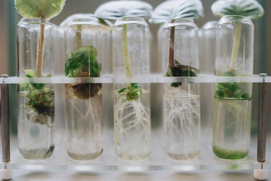
Plant Biology Exercises
Plant biology exercises involve studying photosynthesis, respiration, and anatomical structures. Key experiments include measuring transpiration rates and preparing herbarium sheets to preserve plant specimens for identification.
4.1 Photosynthesis Experiments
Photosynthesis experiments are designed to explore the process by which plants convert light energy into chemical energy; Common experiments include measuring oxygen release using aquatic plants like Elodea or algae. Students often investigate how factors such as light intensity, carbon dioxide levels, and temperature affect photosynthetic rates. Another approach involves using pH indicators to observe changes in dissolved CO2 levels in water. These exercises provide hands-on insights into the light-dependent and light-independent reactions. Controlled environments, such as terrariums, are sometimes used to simulate natural conditions. Data collection and analysis help students understand the relationship between environmental variables and photosynthetic efficiency, reinforcing concepts like the Calvin cycle and energy transfer in ecosystems.
4.2 Preparation of Herbarium Sheets
Preparation of herbarium sheets involves preserving and organizing plant specimens for long-term study. Students typically collect plant samples, ensuring they represent key morphological features. Specimens are cleaned and pressed to remove moisture, often using plant presses or heavy books. Once dry, plants are mounted on sturdy paper or cardboard sheets using glue or tape. Labels are added, detailing species name, collection location, date, and collector’s name. This method allows for detailed observation of plant structures, aiding in taxonomy and identification. Proper storage in a herbarium ensures specimens remain intact for future reference and research.
Animal Physiology Labs
Animal physiology labs provide hands-on experiences in studying organ systems, blood flow, and heart anatomy, offering insights into physiological processes and structure-function relationships while developing practical dissection skills.
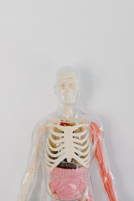
5.1 Dissection and Study of Organ Systems
Dissection and study of organ systems provide a hands-on understanding of anatomy and physiology, allowing students to explore the structure and function of various organs and tissues. This includes examining the heart’s internal anatomy, such as chambers and valves, and tracing blood flow through vessels. Dissections of other systems, like the respiratory or digestive tracts, reveal how organs interact to maintain bodily functions. Proper techniques, such as using scalpels and forceps, are emphasized to ensure accurate observations. These exercises bridge theoretical knowledge with practical application, fostering a deeper appreciation of complex biological systems and their interdependencies.
5.2 Blood Flow and Heart Anatomy
Studying blood flow and heart anatomy involves examining the structural and functional aspects of the cardiovascular system. Students learn to identify chambers, valves, and vessels, tracing blood circulation through the heart. Dissection exercises reveal how blood flows from the atria to ventricles and into arteries. Observing the ventral aorta and its pathways helps understand oxygenated and deoxygenated blood distribution. Proper techniques, such as using scalpels and forceps, ensure accurate dissection. This hands-on approach clarifies how the heart maintains circulatory efficiency, connecting theoretical knowledge with practical observation of cardiovascular physiology.
Molecular Biology Techniques
Molecular biology techniques involve advanced methods like DNA extraction, PCR, and protein analysis, enabling researchers to study genetic material and its functions in-depth for various applications.
6.1 DNA Extraction and PCR Basics
DNA extraction involves isolating genetic material from cells through lysis, centrifugation, and purification. PCR (Polymerase Chain Reaction) amplifies specific DNA sequences using primers and enzymes. These techniques are fundamental in molecular biology for cloning, sequencing, and diagnostic applications. Proper safety protocols and precise temperature control are essential for accurate results. Understanding these methods is crucial for advancing genetic research and biotechnology. Labs often include hands-on practice to master these procedures, ensuring students can apply them in real-world scenarios effectively.
6.2 Protein Analysis and Electrophoresis
Protein analysis involves identifying and studying protein structures, functions, and interactions. Electrophoresis is a key technique used to separate proteins based on size, charge, or other properties. SDS-PAGE (Sodium Dodecyl Sulfate-Polyacrylamide Gel Electrophoresis) is commonly used to denature and separate proteins, followed by staining or Western blotting for detection. These methods are essential in molecular biology for understanding protein expression, diagnosing diseases, and studying enzyme activity. Proper handling of samples and equipment ensures accurate results. Labs often include hands-on practice with electrophoresis to familiarize students with protein analysis techniques and their applications in research and diagnostics.
Ecology and Environmental Studies
Ecology and environmental studies focus on understanding interactions between organisms and their surroundings. Labs include measuring air quality, studying suspended particulate matter, and sampling local flora and fauna. These exercises provide hands-on experience in ecological principles, promoting conservation and sustainability efforts through practical observations and data collection.
7.1 Measuring Air Quality and Suspended Particulate Matter
Measuring air quality involves assessing pollutants like suspended particulate matter (SPM) to understand environmental health. Labs use sensors or filters to collect and analyze air samples, determining SPM concentrations. This helps evaluate pollution levels, identify sources of contamination, and monitor compliance with environmental standards. Students learn to operate equipment, interpret data, and correlate findings with health and ecological impacts. These exercises emphasize the importance of air quality monitoring for public health and sustainability, providing practical insights into environmental science and policy-making.
7.2 Sampling and Identifying Local Flora and Fauna
This exercise involves collecting and identifying plant and animal species in their natural habitats. Students learn to sample flora using methods like pressing for herbarium sheets and fauna through observation or ethical collection. Identification relies on taxonomic keys, field guides, and digital tools. The process emphasizes ecological awareness, biodiversity appreciation, and responsible specimen handling. Data recorded includes species names, habitats, and abundance, fostering skills in ecological surveys and conservation biology. This hands-on approach connects theoretical knowledge with real-world applications in environmental science and species management.
Data Analysis and Presentation
Data analysis involves interpreting experimental results, while presentation includes organizing findings into graphs, charts, and reports. These skills are essential for communicating scientific discoveries effectively and drawing meaningful biological conclusions.
8.1 Graphing and Interpreting Experimental Results
Graphing experimental results is a critical step in data analysis, allowing visualization of trends and patterns. Properly labeled axes, clear legends, and accurate scaling ensure readability. Line graphs, bar charts, and scatter plots are commonly used to represent biological data. Interpreting results involves comparing observed trends to hypotheses, identifying correlations, and assessing statistical significance. Tools like Excel or specialized software facilitate graph creation. Accurate interpretation helps draw meaningful conclusions, while errors in graphing can mislead. Clear communication of results is essential for valid scientific reporting and further analysis in biology experiments.
8.2 Writing Lab Reports and Drawing Conclusions
Writing lab reports involves documenting experimental procedures, results, and conclusions clearly and concisely. Proper structure includes an introduction, materials, procedures, results, and discussion sections. The introduction outlines the experiment’s purpose, while the discussion interprets data, linking results to the hypothesis. Conclusions summarize findings, highlighting their biological significance and implications. Accurate and detailed reporting ensures reproducibility and transparency. Well-organized reports reflect critical thinking and scientific rigor, enabling peers to evaluate the research’s validity and reliability effectively.